Intelligent Arc Handling

ULTRA-HIGH-RESOLUTION IMAGING

UNLOCK THE FULL POTENTIAL OF YOUR ULTRA-HIGH-RESOLUTION CT AND PHOTON-COUNTING CT SYSTEMS


The future of high-resolution CT imaging starts with Dunlee’s extra small focal spots


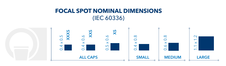
Today’s advanced clinical applications require outstanding spatial resolution for both image quality and workflow. As detector resolution technology continues to push its limits, attention has shifted to increasing spatial resolution via the X-ray source. Dunlee’s small, stable focal spots are engineered to balance optimization of image quality, throughput and heat management to help you get the most out of your ultra-high-resolution CT (UHR-CT) and photon-counting CT (PCCT) systems.


Our Xpert bundles now include three additional extra-small focal spot options, allowing system designers and OEMs to cover the entire spectrum of CT applications with greater flexibility. From our large 1.1 x 1.2 mm focal spot delivering 120 kW for high-throughput imaging, to the smallest 0.4 x 0.5 mm focal spot for ultra-high-resolution imaging, Dunlee’s tubes are designed to support evolving clinical and technical demands.

Advanced Anti-Scatter Technology

Smaller matrix sizes of new high-resolution CT systems demand smaller pixels

THE RISE OF HIGH-RESOLUTION CT IMAGING


CT imaging is undergoing rapid evolution, with spatial resolution and image detail becoming more critical than ever. Two leading approaches are shaping this trend:

Ultra-high-resolution CT (UHR-CT)


Utilizes conventional Energy Integrating Detectors (EID) with reduced pixel sizes for greater resolution.

Photon-counting CT (PCCT)


Incorporates photon-counting detectors that offer both higher resolution through smaller pixel sizes and spectral imaging capabilities.


These technological advances require equally advanced X-ray sources. Smaller detector pixels demand focal spots that are not only smaller, but also thermally and mechanically stable - areas where Dunlee’s technology excels.

CLINICAL APPLICATIONS FOR HIGH-RESOLUTION IMAGING


High-resolution CT applications rely on precise visualization of fine structures, and the demand for improved diagnostic clarity is growing.

Cardiovascular imaging

Cardiology: For visualization of coronary arteries and soft plaques

Characterization of cancer

Oncology: For identification and characterization of small lesions and tumors

Visualization of fine structures in the brain

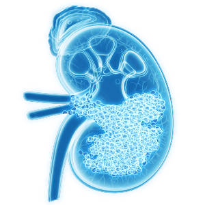
Neurology: For detection of cerebral microbleeds, small infarcts, and evaluation of fine neural structures

Lung visualization

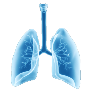
Pulmonology: For detailed lung imaging and detecting interstitial changes

Diagnosis of complex bone pathologies

Orthopedics: For visualization of complex bone pathologies, detection of microfractures, and assessment of implant placement

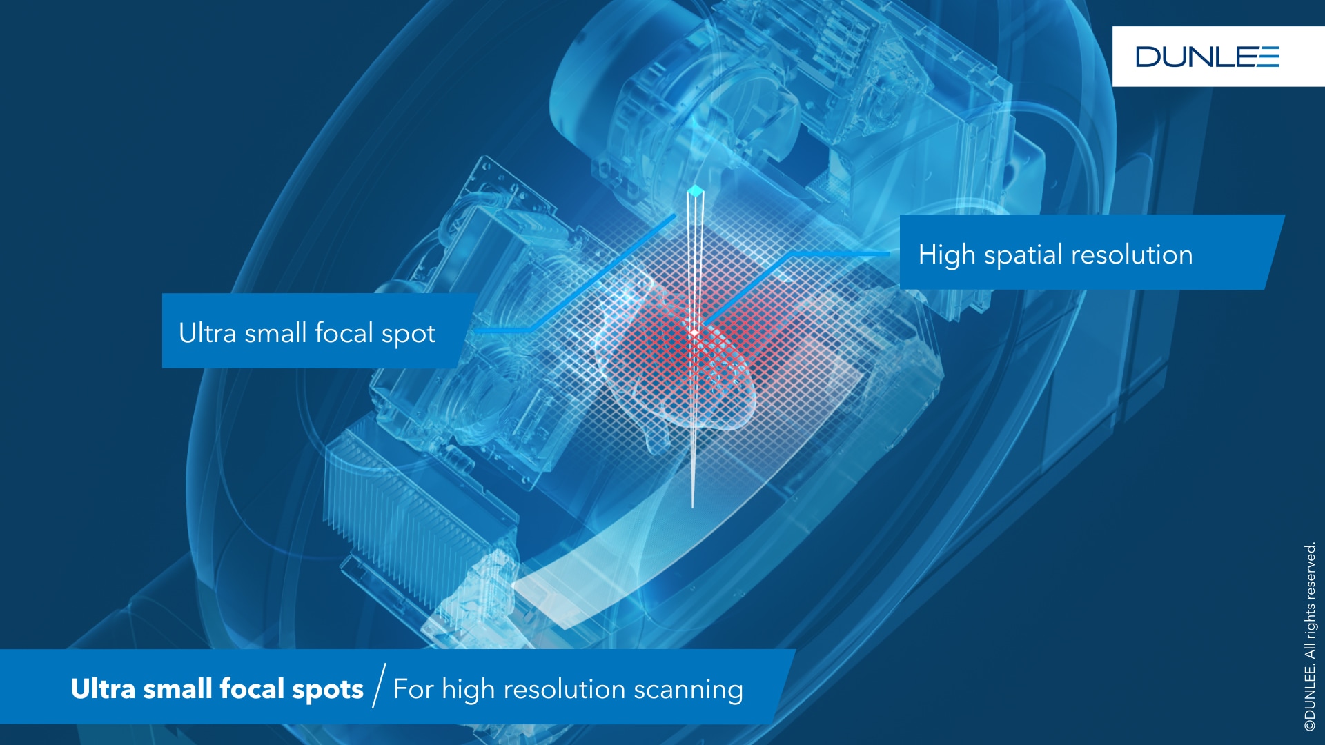
DUNLEE’S ROLE IN ENABLING UHR AND PCCT IMAGING


To fully leverage the capabilities of high-resolution detectors, the performance of the X-ray tube is critical. Our expert engineers have developed small, highly stable focal spots that maintain sufficient power and thermal performance for daily clinical operations.

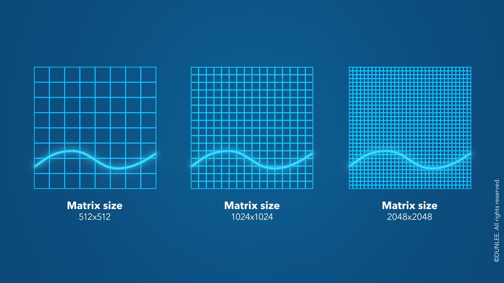
How focal spot size affects CT resolution:

Dunlee XXS Focal Spots

Exploring the relationship between focal spot size & image resolution (measured in LP/mm)

The key physical drivers of spatial resolution include:


  • Detector pixel size: Smaller pixels capture finer detail.
  • Tube focal spot size: Smaller focal spots sharpen projections through reduction of blurring.


However, smaller focal spots increase the power density on the anode, making heat management essential to avoid system downtime. At the same time, high gantry rotation speeds for complex applications - such as heart scans - demand mechanical stability of focal spots, regardless of size.

Three additional focal spot sizes for unlocking the full potential of high-resolution detectors


With the Xpert bundle, Dunlee offers different focal spot options to cover the entire range of CT applications:

Focal Spot Nominal Dimensions

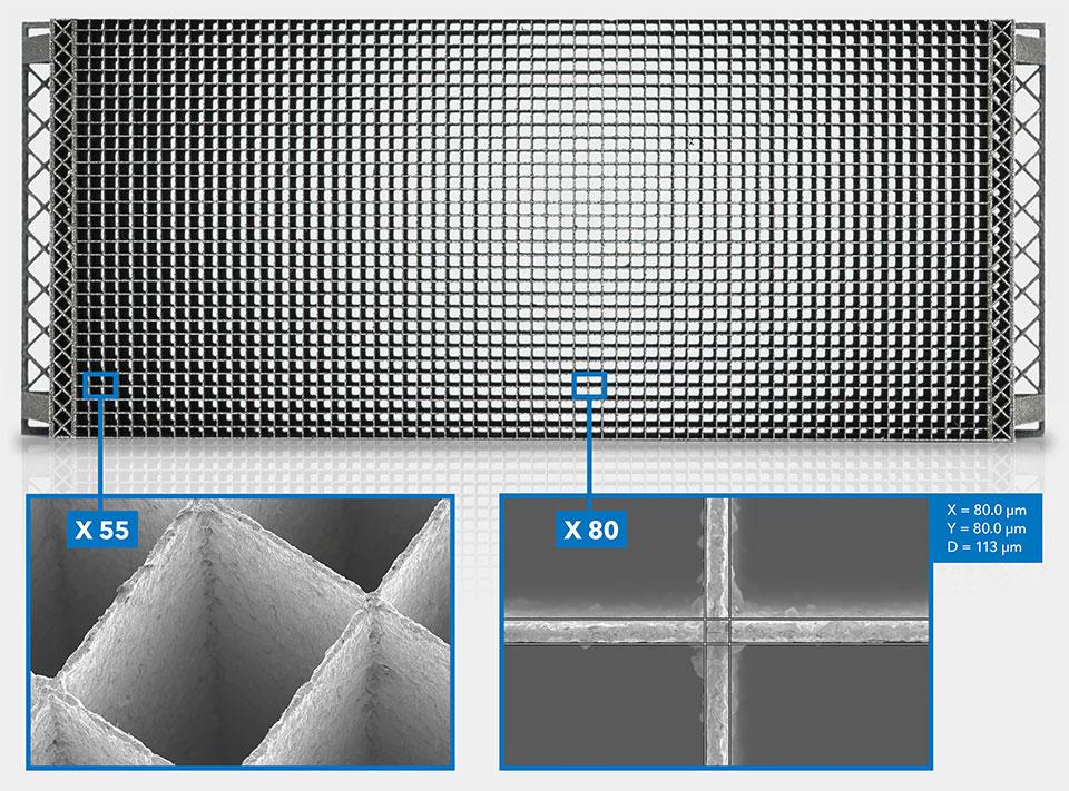
ANTI-SCATTER GRID TECHNOLOGY SUPPORTING IMAGE PRECISION


Advanced CT systems require equally advanced anti-scatter solutions. Dunlee offers a new generation of Anti-Scatter Grids that are optimized for high-resolution imaging:


  • Pure tungsten composition for superior scatter absorption
  • As small as 70 µm feature size with 15 µm precision
  • 3D-printed designs to support emerging UHR and PCCT detector layouts


These grids maintain image sharpness and contrast, particularly when used in systems requiring high precision. To learn more, please visit www.dunlee.com/a-w/3d-metal-printing/our-offer/anti-scatter-grids-for-ct.html.

2D anti-scatter grid

We also support you as you design systems for the next generation of clinical applications.

DUNLEE’S SOLUTION: THE XPERT BUNDLE

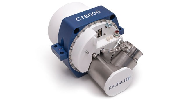
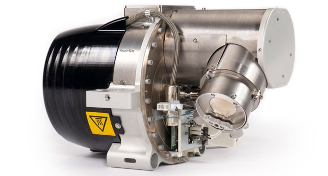
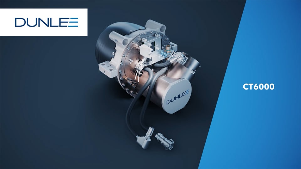
DUNLEE’s Xpert Bundles with CT6000, CT6500 and CT8000 provide extra-small focal spots to address high resolution imaging:

CT6000

CT6500

CT8000

Dunlee CoolGlide LMB

Focal Spot Options
[in mm]

Large: 1.1 x 1.2
Medium: 0.6 x 0.7
Small: 0.4 x 0.7
XS: 0.5 x 0.6
XXS: 0.4 x 0.6
XXXS: 0.4 x 0.5

Large: 1.1 x 1.2
Medium: 0.6 x 0.7
Small: 0.4 x 0.7
XS: 0.5 x 0.6
XXS: 0.4 x 0.6
XXXS: 0.4 x 0.5

Large: 1.1 x 1.2
Medium: 0.6 x 0.8
Small: 0.4 x 0.8
XS: 0.5 x 0.6
XXS: 0.4 x 0.6
XXXS: 0.4 x 0.5

Dunlee CT Bundles

Do you want to learn more about how Dunlee supports system design for high-resolution CT?

From choosing the right focal spot configuration to integration into your imaging system, our experts are here to support you! Reach out to us at marketing@dunlee.com and let’s shape the future of high-resolution CT together.

Sources & Remarks


  1. Gorham, S. et al. Impact of focal spot size on radiologic image quality: A visual grading analysis. Radiography. 2010. 16: 304-313.
  2. Hsieh, S. S. et al. Photon Counting CT: Clinical Applications and Future Developments. IEEE Transactions on Radiation and Plasma Medical Sciences. 2021. 5(4): 441-452. doi:10.1109/trpms.2020.3020212
  3. Xiong, Q. et al. Diagnostic performance of coronary computed tomography angiography stenosis score for coronary stenosis. BMC Medical Imaging. 2024. 24(39).
  4. Hagar, MT et al. Photon-Counting Detector CT: Advances and Clinical Applications in Cardiovascular Imaging. Rofo. 2024. doi: 10.1055/a-2452-0288.
  5. Perera Molligoda Arachchige, A. S. et al. Role of photon-counting computed tomography in pediatric cardiovascular imaging. World Journal of Clinical Pediatrics. 2025. 14(1): 1-8.
  6. Sotoudeh-Paima, S. et al. Photon-counting CT versus conventional CT for COPD quantifications: intra-scanner optimization and inter-scanner assessments using virtual imaging trials. Proc SPIE Int Soc Opt Eng. 2022. doi:10.1117/12.2613003
  7. Kotwani, K. et al. High-Resolution Computed Tomography in the Diagnosis of Temporal Bone Pathologies: A Cross-sectional Study. International Journal of Science and Research. 2024. 13(10): 431-33.
  8. Hsieh, S. S. Overcoming the thermal limits of photon counting CT resolution using focal spot multiplexing: A feasibility study.
  9. Hsieh, S. S. Focal spot rotation for improving CT resolution homogeneity. Proc. SPIE 10573, Medical Imaging. 2018. Physics of Medical Imaging. https://doi.org/10.1117/12.2292472
  10. Shan, Jing, et al. Anode Thermal Analysis of High Power Microfocus CNT X-Ray Tubes for in Vivo Small Animal Imaging. Feb. 2012, pp. 226–34, doi:10.1117/12.911521.
  11. Aurumskjöld, ML et al. A new era of high-resolution CT diagnostics of the lung: improved image quality, detailed morphology, and reduced radiation dose with high-resolution photon-counting CT of the lungs compared to high-resolution energy-integrated CT. Acta Radiol. 2024. 65(10):1211-1221. doi: 10.1177/02841851241269918
  12. Sun, Y. et al. Clinical ultra-high resolution CT scans enabled by using a generative adversarial network. Med Phys. 2023. 50(6):3612-3622. doi: 10.1002/mp.16172.
  13. Sharma, A. et al. A Review of Photon-Counting Computed Tomography (PCCT) in the Diagnosis of Cardiovascular Diseases. Cureus. 2024. 16(11). doi:10.7759/cureus.73119
  14. Milán Vecsey-Nagy et al.Coronary Plaque Quantification with Ultrahigh-Spatial-Resolution Photon-counting Detector CT: Intraindividual Comparison with Energy-integrating Detector CT. Radiology. 2025. 314(3).
  15. Bartlett, DJ et al. High-resolution chest computed tomography imaging of the lungs: impact of 1024 matrix reconstruction and photon-counting detector computed tomography. Investigative Radiology. 2019. 54(3): 129–137
  16. Stayman, JW et al. Multiple focal spots for high resolution CT. Proc. SPIE 12463. Medical Imaging 2023: Physics of Medical Imaging, 124630U(7 April 2023); https://doi.org/10.1117/12.2654455
  17. Henkelman RM. Increased CT tube life. J Comput Assist Tomogr. 1981 Feb;5(1):142-3. doi: 10.1097/00004728-198102000-00028. PMID: 7240489.
  18. Heiner Daerr, D001951504
  19. Zhong, L. F. et al. Measuring Size and Stability of Focal Spot for Linear Accelerator in Industrial CT by Slit Translation Scanning Method. IEEE Transactions on Instrumentation and Measurement, 1. https://doi.org/10.1109/tim.2024.3406806
  20. WHO: Cardiovascular diseases (CVDs)
  21. Kazi, S et al. History Taking, Assessment, and Diagnosis of Patients with Cardiovascular Diseases-Re-defining the Clinical Skills. International Journal of Endorsing Health Science Research. 2022. 10(4): 422–432. https://doi.org/10.29052/IJEHSR.v10.i4.2022.422-432
  22. Bakshi, I. (2024). From Detection to Therapy: The Transformative Power of Radiology in Oncology. International Journal For Multidisciplinary Research, 6(4). https://doi.org/10.36948/ijfmr.2024.v06i04.24949
  23. Graafen D et al. Optimization of the Reconstruction Settings for Low-Dose Ultra-High-Resolution
    Photon-Counting Detector CT of the Lungs. Diagnostics. 2023; 13(23):3522. https://doi.org/10.3390/diagnostics13233522
  24. Seners P, Wouters A, Maïer B, et al. Role of Brain Imaging in the Prediction of Intracerebral Hemorrhage Following Endovascular Therapy for Acute Stroke. Stroke. Published online June 19, 2023
  25. Hildebrand T, Ma Q, Heyward CA, Haugen HJ, Nogueira LP. Advanced soft tissue visualization in conjunction with bone structures using contrast-enhanced micro-CT. Published online October 17, 2024. doi:10.1117/12.3027063
  26. Lee SK, Joo MW, Kim JY, Bernthal NM. Postoperative Imaging of Bone and Soft Tissue Tumors in the Extremity: A Comprehensive Review. Diagnostics. 2024;14(24):2794. doi:10.3390/diagnostics14242794
  27. Vittorio Di Trapani, Francesco Brun. Pre- and post-reconstruction digital image processing solutions for computed tomography with spectral photon counting detectors. Nuclear Instruments and Methods in Physics Research Section A: Accelerators, Spectrometers, Detectors and Associated Equipment. 2021. 1010. https://doi.org/10.1016/j.nima.2021.165510.
  28. McCollough, C.H., Rajendran, K., Leng, S. et al. The technical development of photon-counting detector CT. Eur Radiol 33, 5321–5330 (2023). https://doi.org/10.1007/s00330-023-09545-9
  29. Yamaguchi I, Morimoto A. Influence of rotation speed of X-ray computed tomography on image quality. Nihon Hoshasen Gijutsu Gakkai Zasshi. 2004 Jan;60(1):79-86. Japanese. doi: 10.6009/jjrt.kj00000922261. PMID: 15041910.
  30. Khan AA, Labbe JC. Advanced ceramic matrix composites for high energy x-ray generation. Adv. Nat. Sci.: Nanosci. Nanotechnol. 2 (2011) 045015 (8pp).


Click here to read moreClick here to read less

Dunlee Logo

About Dunlee

Dunlee is a brand of the Philips Company Group and distributes all products, solutions and services for the third-party imaging business. The product portfolio includes CT, MR and X-ray solutions for OEMs and 3D printed tungsten products.

© 2017 - 2025 Koninklijke Philips N.V.  All rights reserved.

You are about to visit a Philips global content page

Continue

Our site can best be viewed with the latest version of Microsoft Edge, Google Chrome or Firefox.